If you’ve booked a first orthodontic consultation, you may be wondering why the appointment involves more than someone just looking at your teeth. X-rays and digital scans are a standard part of the process, and they exist for reasons that go well beyond checking how straight your smile looks.
A Visual Exam Only Tells Part of the Story
Looking at teeth from the outside shows crowding, spacing, and obvious bite issues. It does not show what’s happening below the gumline. Root positions, bone levels, and the relationship between the jaws are only visible with imaging, and all three factor into how a treatment plan gets built.
A dentist or orthodontist can tell a lot from a smile at first glance, but treatment planning is different from a casual observation. Two people can have teeth that look similarly crowded and end up with very different treatment plans once the imaging comes back.
What X-Rays Reveal
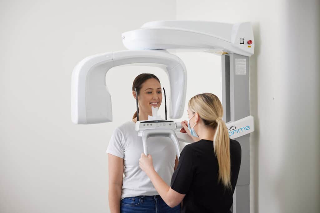
A panoramic X-ray gives a wide view of the entire mouth in a single image. This includes the position of unerupted teeth, wisdom teeth development, and any extra or missing teeth that wouldn’t be obvious otherwise. It also shows root angles, which matter because moving a tooth with a tilted root requires a different approach than moving one with a straight root.
Cephalometric X-rays, which capture the side profile of the skull, help assess how the upper and lower jaws relate to each other. This is particularly useful for evaluating bite issues like an overbite or underbite, since jaw position sometimes plays a bigger role than tooth position alone.
These two types of imaging work together rather than replacing one another. The panoramic view maps out individual teeth and roots, while the cephalometric view maps the broader skeletal relationship those teeth sit within.
What Digital Scans Add
A digital scan, taken with an intraoral scanner like iTero, maps the exact shape and position of every tooth in great detail. Unlike a physical impression, a digital scan is quick and doesn’t involve biting into a tray of putty. It also creates a 3D model that can be used to simulate how teeth might move over the course of treatment.
Digital scans make it easier to track changes over time as well. Comparing a scan taken today to one taken a year from now shows precisely how much movement has occurred, which helps confirm that treatment is progressing as expected.
For patients who are curious what treatment might actually look like, some digital scan systems can generate a simulation showing an approximate progression of tooth movement. This isn’t a guarantee of the final result, but it gives a general sense of the plan before any brackets or aligners are involved.
Why This Information Changes the Treatment Plan
Catching Problems Early
Imaging sometimes reveals things a visual exam would miss entirely, such as an impacted tooth that hasn’t broken through the gum yet, or a wisdom tooth positioned in a way that could affect nearby teeth. Identifying these issues before treatment starts allows the plan to account for them from day one instead of adjusting course partway through.
Choosing Between Treatment Options
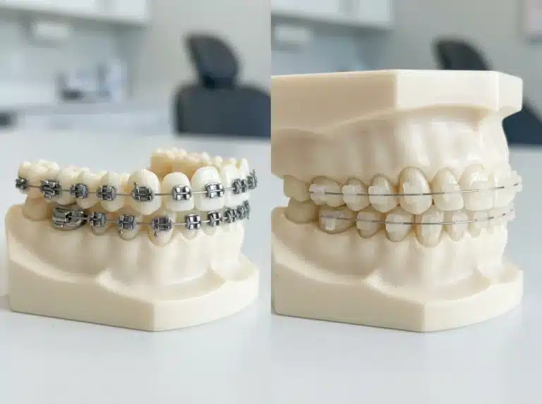
Whether a patient is a better fit for traditional braces, clear braces, or Invisalign often depends on details that only show up in imaging. Root length, bone density, and the position of the bite all influence which approach will work best for a particular mouth, not just what looks acceptable in the mirror.
Setting Realistic Expectations
Because imaging shows the full picture, it also helps set an accurate timeline. Two patients with similar-looking crowding from the outside can have very different root and bone situations underneath, which means their treatment length may differ even if their starting smiles look alike.
This is also why timelines given at a consultation are estimates rather than fixed promises. Treatment can move faster or slower than expected depending on how the teeth and bone respond, which is something imaging helps predict but can’t determine with total certainty.
Is the Imaging Safe?
Digital X-rays used in orthodontic offices involve a very low dose of radiation, and digital sensors require significantly less exposure than older film-based X-rays did. Lead aprons and thyroid collars are used as an added precaution during any X-ray capture. Digital scans involve no radiation at all, since they rely on a small handheld camera moved gently around the mouth.
Parents bringing in younger children sometimes ask specifically about radiation exposure. It’s a fair question, and the honest answer is that a panoramic X-ray involves less radiation than many people expect, roughly comparable to a few hours of everyday background exposure.
What to Expect During Your Consultation
Before the Scan
Most consultations start with a conversation about your concerns and goals, followed by a visual exam. From there, the imaging portion typically takes only a few minutes for both the X-rays and the digital scan combined.
After the Scan
Once the images are captured, they’re reviewed together with the visual findings to put together a treatment recommendation. Patients usually get to see their own scan on screen, which makes it easier to understand exactly what’s being addressed and why. Questions about the images are welcome at this stage, since seeing the reasoning behind a plan tends to make the whole process feel less abstract.
Book Your Consultation With Kelowna Orthodontics
Understanding what’s happening beneath the surface is the first real step toward a treatment plan that fits your mouth specifically, not a generic approach. Kelowna Orthodontics uses digital X-rays and iTero scanning at every consultation to build a plan based on your actual anatomy.
Book a consultation with Kelowna Orthodontics
- Call (250) 763-3312 to ask about your first visit
- Learn more about Invisalign options
- Request a free consult, no referral needed
- Visit the Kelowna Orthodontics office on Harvey Ave for a digital scan